
Differential Interference Contrast Image Gallery
Spirogyra Filamentous Algae
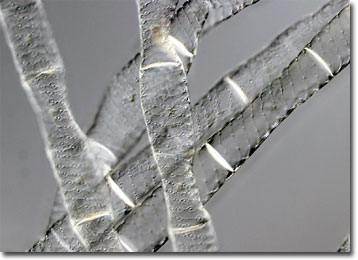
Though many refer to filamentous algae belonging to the genus Spirogyra as pond scum, the delicate green strands are sometimes known by more complimentary terms, such as water-silk or mermaid’s tresses. The beauty of Spirogyra is most prominent under the microscope, where helices of emerald-hued chloroplasts can be seen spiraling the length of its tubular cells.
More than 400 species of Spirogyra are known to be in existence and most are found free-floating in freshwater environments. The algae are particularly abundant in nutrient-rich environments and large populations can be an indication of over-fertilization caused by contaminated storm water. At night or when it is overcast, it may be difficult for the casual observer to determine the level of algae in a pond or other body of water. However, on sunny days, oxygen bubbles produced during photosynthesis cause Spirogyra to rise to the surface of the water, often in thick, tangled mats.
The reproductive process of Spirogyra is usually asexual, individual strands breaking off to form new strands. In unfavorable conditions, however, the algae may switch to a sexual means of proliferation. Adjacent strands are capable of extending temporary conjugation tubes toward one another in order to exchange the necessary genetic information to form a zygospore. On rare occasions, cells of the same filament perform a similar reproductive action. The cell wall of the resultant zygospore is thick and resilient, which allows the organism to survive until environmental circumstances improve.