
Differential Interference Contrast Image Gallery
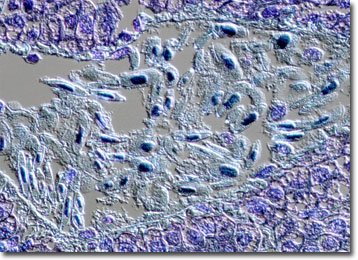
Nucleic Acid Stains
The first synthetic dye, aniline purple, was developed in 1856 by English chemist William Perkin. Though other synthetic chemists soon replicated the feat, it was at least another twenty years before using dyes and stains became common use in most scientific laboratories.
In modern times, the process of staining cells to observe their features is widespread, and scientists have an array of stains, generally composed of heterocyclic organic dyes, to choose from. In order to make a suitable stain selection, the chemical and electronic properties of the dye molecules with respect to their interaction with cellular components must be considered. For instance, stains that have a net positive charge in aqueous or buffered solutions have an affinity for negatively charged biological structures and selectively bind to them. Hydrophilic stains, on the other hand, tend to stain hydrated biological bodies, such as the exterior of proteins and nucleic acids. Contrariwise, stains hydrophobic in nature concentrate on membranes, lipids, and the interior of proteins.
Ethidium bromide is a typical nucleic acid stain used by molecular biologists. Dark red and in crystal form when not in an aqueous solution, the fluorochrome exhibits a reddish-orange fluorescence that can be detected under both long and short ultraviolet wavelengths of light. Furthermore, ethidium bromide is a relatively large, flat basic molecule that somewhat resembles a DNA base pair and because of its structure can easily insert into a DNA strand. Thus, the substance is extremely useful for experimental purposes, though it can dangerous to handle and should be employed with caution.