
Differential Interference Contrast Image Gallery
Mouse Kidney Thin Section
Since it is often not possible to study human organs, scientists frequently must depend upon laboratory animals for their research purposes. One of the most popular of all laboratory animals is the mouse, which has a long history of helping humans understand a wide variety of physical conditions, diseases, and other phenomena.
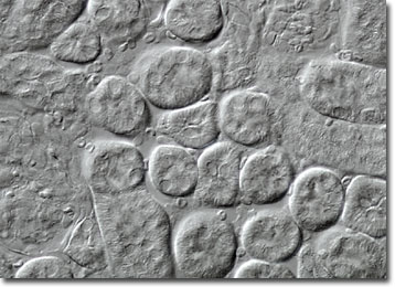
The close mammalian relationship between mice and humans means that many of their organs function in the same way, even though they may vary in size or location. The kidneys, for instance, maintain water balance and expel metabolic wastes in both species. They also have a similar appearance; bean shaped, brownish red, and granular. The outer region of both human and mouse kidneys is called the cortex and the inner area is known as the medulla. Nephrons, which are the functional units of the kidneys, stretch across both sections and are responsible for filtering blood, reabsorbing water and nutrients, and secreting wastes.
Due to their similarity to human kidneys, mouse kidneys are extremely useful in scientific studies. For example, researchers have gained a much better understanding of many common forms of kidney disease by using genetically engineered mice as models. Moreover, recently a team of scientists working at the Institute of Science successfully used human stem cells to grow new organs inside of kidney deficient mice. The miniature human kidneys were fully functional and were carefully monitored in order to gauge the possibility of rejection. Further research may make it possible for humans to grow their own replacement kidneys, offering new hope to the tens of thousands of people on waiting lists for organ donations.