
Differential Interference Contrast Image Gallery
Desmid Algae
Desmids are typically unicellular green algae that proliferate in freshwater environments, including ponds, swamps, and bogs. Similar, in a sense, to snowflakes, it is difficult to find two identical desmids because they exist in a tremendous variety of shapes and sizes.
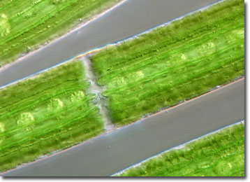
Since they are so small, the beauty of desmids tends to go unnoticed unless they are examined through a microscope. Strikingly symmetrical, each individual desmid is divided into two mirror-image halves, or semicells, which are connected by an isthmus that contains the nucleus. The semicells may be globular, disc, spindle, or crescent shaped, and can feature a cell wall ornamented with spines or spectacular patterns of granules and tubercles. Desmids have an even more unusual appearance when they are in the process of dividing. During this time, two new semicells are created in between the old semicells, often remaining attached for a short period.
The increasing incidence of fertilizer run-off in still bodies of water has been detrimental to desmid populations. The organisms cannot reproduce as quickly as many other forms of algae in nutrient-rich environments and tend to diminish when placed in a situation of heavy competition. Thus, due to their dependence on clear, unadulterated water, desmids have become rare in many parts of the world. However, as a consequence of their stringent environmental demands, the organisms are very useful in monitoring the conditions of aquatic habitats.