
Differential Interference Contrast Image Gallery
Cucumber Tapeworm (Dipylidium caninum)
Scientifically described as Dipylidium caninum, the cucumber tapeworm gained its common name from the characteristic appearance of the segment-like proglottids that it sheds. Found throughout the world, the parasite is most common in areas with high populations of domestic dogs and cats.
Proglottids, which are actually closer in size and shape to grains of rice, are a primary indicator of cucumber tapeworm infection. They contain egg cases and may be found in the bodily waste of definitive hosts, such as canine and feline species and, occasionally, humans. Definitive host infection occurs through the accidental ingestion of a contaminated intermediate host, which may be either a flea or a louse. Adult lice and larval fleas become infected by swallowing eggs released from the proglottids of a mature cucumber tapeworm.
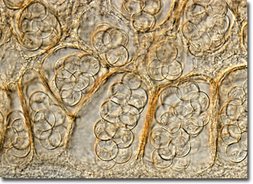
In order to attach themselves firmly to the wall of a host’s small intestine, each adult cucumber tapeworm features a scolex with four suckers and a retractable projection known as the rostellum that is lined with several rows of hooks. Once they reach their final destination, individual worms may grow to substantial sizes, sometimes achieving lengths of up to 50 centimeters. When sexual maturity is achieved, the hermaphroditic parasites are capable of fertilizing their own eggs. The eggs of cucumber tapeworms are each about 50 microns in diameter and are released in cases that may hold up to thirty organisms.