
Differential Interference Contrast Image Gallery
Commercial Sponge Fibers
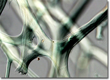
The soft, elastic skeletal systems of such sponge species as Spongia officinalis and Spongia graminea have resulted in their commercial exploitation for thousands of years. The relatively recent advent of synthetic sponge materials has, however, greatly decreased modern utilization of natural varieties.
The use of sponges has been prolific and varied throughout history. In ancient times, Greeks and Romans commonly utilized them for cleaning, bathing, and painting, and soldiers of those civilizations used them to hold drinking water. The ancient Chinese, on the other hand, burned sponges as a treatment for goiter and other medical problems, a technique that was also carried out in Europe during the Middle Ages. Sponges are often utilized in modern medical practice, as well, but primarily as an as an absorbent surgical material.
Sponges do not naturally exhibit the fresh, golden appearance that they have when modern consumers buy them from stores. In fact, when divers first bring them up from the sea they are black and rather unpleasant. A systematic process of washing and pressing the sponges must be followed in order to break down their external membrane and tissues. When only the skeletal fibers of the creatures remain, they are trimmed, dried, and frequently immersed in a mixture of water and hydrochloric acid, a procedure that gives them a blonde coloring, before being sold by merchants.