
Differential Interference Contrast Image Gallery
Stained Chinese Liver Fluke Eggs
The Chinese, or Oriental, liver fluke is a parasitic flatworm that most often proliferates in Asian countries. Humans primarily act as definitive hosts of the trematodes, but other animals may also be infected.
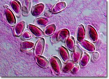
Adult members of the species reside in the bile ducts of the liver, generally growing to lengths of approximately one inch. They may live for more than 20 years and can produce a prolific number of eggs, as many as 4,000 in a single day. The eggs have a characteristic flask-like shape that features a prominent operculum at one end and a spine at the opposite extremity. They are passed through the intestines and are voided from the body with feces. Though the miracidium held inside the eggs is already well developed at this point, it does not hatch until it is ingested by an intermediate snail host. When their gestation is complete, the newly formed trematodes exit the snail and burrow into fish, where they encyst inside muscle tissue.
Humans and other animals become infested with the Chinese liver fluke by consuming raw or undercooked fish that are contaminated with the parasites. Thus, it is in areas where cooking seafood is uncommon that the highest rates of infection exist. Currently, millions of people are believed to suffer from Chinese liver fluke infection, known as clonorchiasis, but with varying degrees of symptoms. Abdominal pain and digestive problems are fairly frequent occurrences, but only severe cases are accompanied by jaundice, fibrosis, and enlargement of the liver. The most common method of treating those that have been diagnosed with clonorchiasis is administration of Praziquantel but, as the saying goes, prevention is the best cure.