
Differential Interference Contrast Image Gallery
Lancelet (Amphioxus) Pharynx
The lancelet, or amphioxus, is a diminutive marine organism that is similar in appearance to an eel. Members of the phylum Chordata, lancelets do not often reach lengths of more than three inches and lack eyes, as well as a distinct heads.
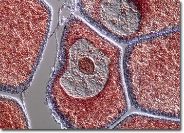
Though capable of swimming, lancelets typically remain buried in the sand or mud that lines the ocean floors. To feed, they extend the anterior portion of their bodies upward out of the muck and the cilia that line their gill slits direct water toward their mouth openings. From the mouth, the water moves into the pharynx, the mucous membrane of the gill basket capturing edible organisms in the water and passing them onto the gut, where digestion begins with the aid of various enzymes. Unlike other chordates, lancelets are capable of phagocytosis, a digestive process in which individual cells consume food particles.
Lancelets breed multiple times each year in tropical regions of the world, but only once in temperate zones. Thus, large populations of the aquatic creatures frequently develop in warm coastal waters, a benefit for the environment in those areas due to their filtration capabilities. However, in addition to creating clean water, the abundant lancelets have other ways of making themselves useful to humans. For instance, in some areas, such as along sections of the coast in China, lancelets have become an integral part of the fishing industry.