
Smith, Beck & Beck Large Best or No. 1
Smith & Beck created a style of compound microscope dubbed the Large Best or No. 1 Stand, and set a design trend with the brass scientific instrument that lasted more than 20 years. The binocular model illustrated below was redrawn from photographs of the original microscope, which was described by Gerard Turner in his book The Great Age of the Microscope.
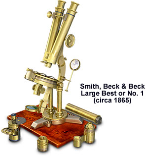
The microscope stand, which was dated 1855 based on its serial number, is signed "Beck & Beck". According to Turner's notes, however, the binocular microscope must have been reconditioned in the Smith, Beck & Beck workshop based on a pair of 1861 Wenham body tubes and the case with accessories that was illustrated first in Richard Beck's 1865 book, A Treatise on the Construction, Proper Use, and Capabilities of Smith, Beck, and Beck's Achromatic Microscope. The three achromatic lenses are not signed, but the storage can for the 1/20-inch objective bears the inscription "Smith, Beck & Beck, 31, Cornhill, London," which dates it after 1857 when the firm changed names.
The inscription "Smith & Beck 6, Coleman St, London" identifies the pre-1857 storage tins for the 2/3-inch and 1/8-inch achromat objectives. Two pairs of matched eyepieces and an erector lens system complement the optical system. A pair of Lieberkuhn reflectors and an epi-illuminator, which is numbered and signed "Smith Beck and Beck Patent London," allow normal viewing of opaque specimens under oblique or epi-illumination. The relatively simple, but innovative illumination system, which facilitates reflective light microscopy, consists of a cylinder screwing into the nosepiece, and incorporates a thin glass disc on a turn-pin.
Coarse focus is achieved with the body tubes racked in a Jackson-type groove, while the fine focus mechanism is controlled by a lever that is mounted on the nosepiece. The Wenham binocular tubes may be exchanged for a draw tube (illustrated at the rear of the mahogany base board). Featuring a six-inch engraved scale, the monocular draw tube can host the plano-concave mirror, which is held by an articulated arm, which slides on a collar mount. On the lower limb, a Wenham parabolic condenser (not mounted in the illustration) holds an adjustable central stop, an Amici prism, or a Nicol prism polarizer.
Mounted on a sliding bar, a second Amici prism enables the microscopist to examine specimens under oblique illumination. The precision optical instrument is fitted for a choice of three analyzers that mount in the ocular lens systems, including two with Nicol prisms and one with a tourmaline plate. A cut-away side of the condenser accommodates three, rotating selenite crystals that are mounted on a dovetailed bracket, while an iris diaphragm that is signed "R. & J. Beck, London" was added at a later date. To add to the versatility of the precision optical instrument, the updated brass binocular microscope features a mechanical stage that is installed over a rotating plate. The stage is turned by a pinion and is divided into degrees that are engraved in the brass. An aperture disc with nine positions is incorporated into the socket for the condenser, thus providing the microscopist more control over illumination, specimen contrast, and resolution with the modified microscope.
BACK TO NINETEENTH CENTURY MICROSCOPES