
French Compound Monocular Brass Microscope
Built sometime after 1875, this brass microscope was offered to customers with either an oblong or oval base and was moderately priced for the period. The model featured below was redrawn from photographs of the original microscope, which is part of the Billings microscope collection at Walter Reed Army Hospital in Washington DC.
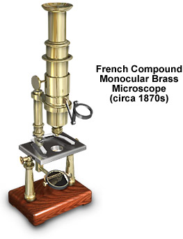
Illustrated with a wooden base, the microscope is supported by two tubular pillars that are approximately two inches high. Attached to the pillars is a stage brass stage plate having a collar and flanges made with the same casting. The tubular limb is attached to the stage collar, and protrudes beneath the stage to support a gimbal with a double mirror to illuminate the specimen.
A U-shaped slide holder is positioned above the stage and secures specimens through tension on the glass microscope slide. The limb also supports a small arm with a double milled-head pinion to which the body tube is attached. On the front of the arm is a universal swivel-joint hinge that holds a condensing lens. The body tube is fashioned from brass and has a short cone-shaped nose and a drawtube with a field lens. Focus is achieved with a rack mechanism that translates the drawtube within the body tube. A single eye lens is mounted in a brass housing and screws into the top of the body tube.
BACK TO NINETEENTH CENTURY MICROSCOPES