
John Dollond Compound Monocular Microscope
This early nineteenth century Dollond microscope was considered to be among the finest optical instruments developed prior to the introduction of achromatic microscope objectives. The model featured below was redrawn from photographs of the original microscope, which is part of the Billings microscope collection at Walter Reed Army Hospital in Washington DC.
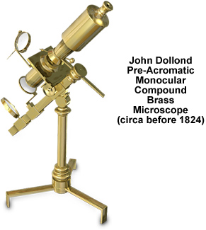
Exhibiting excellent workmanship, the compound monocular microscope is similar in design and execution to the "Jones Most Improved" instrument series and is signed on the base, "Dollond, London". A folding tripod base anchors the circular pillar that is surmounted by a compass joint. Affixed to the joint is a short arm that suspends a limb measuring 10-1/2-inches in length. A double mirror (2-inches in diameter) is clamped onto a sleeve positioned at the lower end of the limb.
Illumination is concentrated by a 1-1/2-inch diameter condenser secured on an adjustable angled limb located above the mirror. The body tube affixes at the nose to the upper portion of the limb and remains stationary. There is no drawtube and focus is accomplished by adjusting the stage position in relation to that of the objective by turning a pinion located on the stage sleeve. Additional optical features include an eyepiece that is comprised of three lenses and a field lens.
BACK TO NINETEENTH CENTURY MICROSCOPES