
Bertrand Box Microscope
Meticulously crafted in Paris, France, the miniature Bertrand drum microscope is accompanied by a beautiful mahogany box and dates back to about 1841. The model featured below was redrawn from photographs of the original microscope, which is part of the Billings microscope collection at Walter Reed Army Hospital in Washington DC.
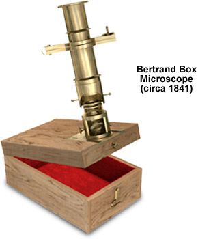
The mahogany storage box is an integral part of the microscope and is designed to serve as a base that anchors the barrel-shaped instrument. Comprised of individual units that are detachable, the brass monocular microscope is surprisingly compact -- the storage box measures only 2-1/2 x 3-7/8 x 1-5/8 inches. Fine focus is achieved by sliding the body tube closer to or farther away from the specimen. The mechanism for fine adjustment is composed of a steel spring clamp positioned above the cylinder assembly and is manipulated by turning a milled-head screw. Focusing is further enhanced by a choice of three interchangeable objectives of varying magnification. The specimen stage of the compound monocular microscope is composed a single fixed plate. Although fitted with a 7/16-inch adjustable mirror, uniform illumination can only be minimally achieved because the instrument lacks a substage condenser.
BACK TO NINETEENTH CENTURY MICROSCOPES