
Differential Interference Contrast
Interactive Tutorials
Optical Sectioning with Phase Contrast and DIC
One of the primary advantages of differential interference contrast (DIC) microscopy over phase contrast is the ability to utilize the instrument at full numerical aperture without suffering the masking effects of phase plates or condenser annuli, which severely restrict the size of the condenser and objective apertures. The major benefit is improved axial resolution, particular with respect to the ability of the DIC microscope to produce excellent high-resolution images at large aperture sizes. This interactive tutorial explores and compares optical sectioning of thick specimens with DIC and phase contrast, and reveals the benefits of unrestricted aperture effects on obtaining well-defined sections.
The tutorial initializes with the image of a randomly selected specimen appearing in each of the Specimen Image windows. The left-hand window, entitled DIC Specimen Image, contains an image of the specimen captured with a differential interference contrast optical system, while the right-hand window (Phase Contrast Specimen Image) displays the same specimen when imaged in phase contrast. The microscope focal plane displayed in the twin windows is identical for each specimen. In order to operate the tutorial, use the Objective Focus slider to simulate the effects of simultaneously imaging the specimens through various axial focal planes. As the slider is translated to the left, the upper focal planes become visible, and moving the slider to the right reveals the lower focal planes. A new specimen can be selected at any point by using the list available in the Choose A Specimen pull-down menu.
Because phase contrast images can become ambiguous (or even experience a reversal of contrast) when the specimen optical path length fluctuates over a large range, the technique should be restricted to specimens having a path difference of one-tenth wavelength or less, as previously discussed. In other words, thick specimens, or those having wedge-shaped structures, are generally not suitable for quantitative investigations with phase contrast microscopy. Thin specimens also tend to produce superior images in differential interference contrast, but thicker specimens (having an optical path difference between one-tenth and a full wavelength) can also be imaged at wide apertures using optical sectioning techniques.
The illumination aperture in phase contrast is fixed by the size of the condenser annulus and cannot be varied to achieve increased or decreased depth of field. This restriction hampers observations of specimens containing significant halo artifacts, which can sometimes obscure focused specimen features due to overlapping details originating from excessive light originating outside the objective focal plane. Such problems often arise when attempting to image specimens that are too thick for phase contrast. However, as previously discussed, the optical sectioning capability of differential interference contrast microscopy (at wide condenser aperture sizes) enables excellent images to be obtained with relatively thick specimens.
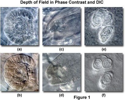
The benefits of large aperture optical sections from thick specimens in differential interference contrast is illustrated in Figure 1, and compared to the same viewfields imaged with phase contrast. Figure 1(b) illustrates a medusa bud from a reproductive polyp of the Obelia hydrozoan imaged at high magnification in DIC with the condenser iris diaphragm opening set to approximately 95 percent of the objective aperture size. The resulting thin optical section reveals sharply focused image details of the medusa features, which are only minimally obscured by light originating away from the focal plane. When the same viewfield is examined in phase contrast (Figure 1(a)), the medusa image is blurred by halos and exhibits reduced resolution due to the restricted aperture of the optical system.
Similar situations are presented in Figures 1(c) through 1(f). Figure 1(c) depicts overlapping wing scales from the monarch butterfly (Danaus plexippus), one of the most common butterflies in North America. Individual scales are not distinguishable in Figure 1(c) because of halo artifacts and indistinct features that are not in the plane of focus. In general, the image is confused by numerous artifacts, making specimen features difficult to decipher. Imaging the same specimen in differential interference contrast (Figure 1(d)) eliminates many of the distracting artifacts positioned away from the focal plane and clearly delineates the surface features in a single wing scale. Likewise, an egg packet within the abdomen of a canine cucumber tapeworm (Dipylidium caninum) reveals sharp edges surrounding individual eggs when imaged in DIC (Figure 1(f)). However, the focused eggs are devoid of distinctive structure in phase contrast (Figure 1(e)), primarily due to halos from others eggs situated away from the focal plane.
The optical sectioning capability of differential interference contrast at large condenser aperture sizes and high numerical aperture is critical for imaging single focal planes in thick phase specimens. Details and stray light from features lying outside of the immediate focal plane do not comprise DIC images in the same manner as phase contrast images, which are often complicated by halo artifacts. As a result, differential interference contrast can be employed to obtain excellent images under somewhat unfavorable conditions (very thick specimens), even when large depth of field renders the identification of phase structures impossible in phase contrast microscopy due to overlapping details.
Contributing Authors
Jan Hinsch - Leica Microsystems, Inc., 90 Boroline Road, Allendale, New Jersey, 07401.
Matthew J. Parry-Hill and Michael W. Davidson - National High Magnetic Field Laboratory, 1800 East Paul Dirac Dr., The Florida State University, Tallahassee, Florida, 32310.
BACK TO DIFFERENTIAL INTERFERENCE CONTRAST MICROSCOPY