Brightfield Microscopy Digital Image Gallery
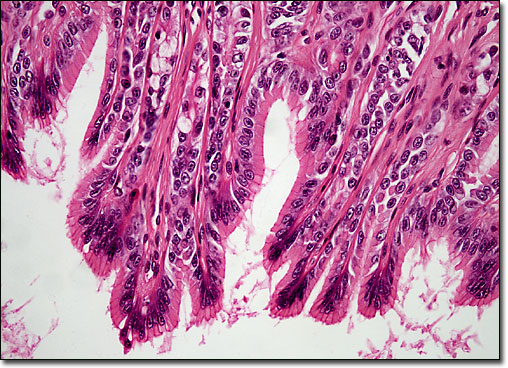
Simple Columnar Epithelium
|
Columnar epithelium can be found along the intestinal treact, spanning from the end of the esophagus to the rectum. This type of tissue also occurs in the ducts of various glands. Epithelial cells are taller than they are wide and contain nuclei along their bases. The membranes that surround them are relatively thin, but can be easily viewed with the aid of a microscope. One of the best-known examples of columnar epithelial cells appears as a covering of the projections called villi found in the small intestine. The functions of these and other cells of the epithelium are various, but essentially involve absorption, secretion, and protection. |
© 1995-2022 by Michael W. Davidson and The Florida State University. All Rights Reserved. No images, graphics, software, scripts, or applets may be reproduced or used in any manner without permission from the copyright holders. Use of this website means you agree to all of the Legal Terms and Conditions set forth by the owners.
This website is maintained by our
|