
Brightfield Microscopy Digital Image Gallery
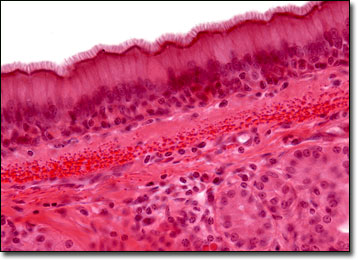
Pseudostratified Ciliated Columnar Epithelium
The cells that comprise the epithelial membranes are variously shaped and are named accordingly. As implied by their moniker, columnar epithelial cells are taller than they are wide, appearing like numerous miniature pillars.
The cells of the epithelium are tightly packed, providing protection to surfaces of the body that are closest to the exterior world. The cells may also be organized into numerous layers, depending upon their location within the body. In such instances, a number of different types of epithelium may be identified, such as stratified and transitional epithelium. The epithelium of the skin is perhaps the best example of the stratified type of the tissue. Within the epithelium of the skin, three different layers of cells exist, each exhibiting a different shape. The multiple layers are necessary in such a location because exposure to the outside world results in the outer layer of cells being continuously shed. Thus, the deepest layer of epithelial cells found in the skin are formative, and the middle layer is an intermediate location where the newly produced cells gradually transform until they must replace the surface cells.
Unlike the epithelium of the skin, a pseudostratified ciliated columnar epithelium appears to have multiple layers, but is actually only comprised of a single sheet of cells. The positioning of the nuclei within the individual columnar cells causes this illusion. These structures, which are easily identifiable with the help of a microscope, are found at various levels, creating a stratified appearance. A microscope also facilitates the observation of the tiny hairlike cilia that line the cells. Found most heavily along the respiratory tract, pseudostratified ciliated columnar epithelial cells help trap and transport particles brought in through the nasal passages and lungs.
BACK TO THE BRIGHTFIELD MICROSCOPY IMAGE GALLERY