
Brightfield Microscopy Digital Image Gallery
Oleander Leaf
Oleanders are evergreen shrubs of the genus Nerium that belong to the dogbane family, Apocynaceae. Believed to be native to the Mediterranean region, these beautiful, but deadly organisms, were well known to both the ancient Greeks and Romans.
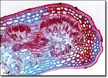
Though the appearance of oleanders varies by species, most exhibit thick, lance-like leaves and beautiful flowers that develop in clusters. These flowers generally bloom in the spring and may appear in a variety of colors, including pink, red, yellow, apricot, and white. Hardy and fast growing, oleanders are widely utilized in landscaping, especially in cities and along highway medians, despite the fact that they are extremely poisonous. The toxin present in oleanders is most concentrated in the shrub’s sticky, milky sap, but is contained in smaller quantities in all its many parts. Thus, no part of an oleander should ever be consumed and even contact should be avoided since its toxin can cause serious skin irritation.
In the 1990s, a plant disease lethal to oleanders was discovered in southern California. Dubbed oleander leaf scorch, this malady is caused by the bacterium Xylella fastidiosa, which is a different strain of the species that causes almond leaf scorch and Pierce's disease in grapevines. Symptoms of oleander leaf scorch, which initially include the drooping and yellowing of leaves on one or more branches, are often mistakenly believed to be brought on by drought. However, shrubs suffering from the disease will not recover when watered since the presence of bacteria limit the flow of water. Instead, the symptoms spread to more branches and the plant eventually dies, usually within 3 to 5 years of the earliest signs of disease.
BACK TO THE BRIGHTFIELD MICROSCOPY IMAGE GALLERY