
Brightfield Microscopy Digital Image Gallery
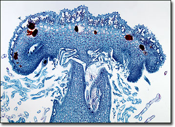
Marchantia Liverwort Archegoniophore
Liverworts are spore-producing plants that belong to the class Marchantiopsida of the division Bryophyta, which also contains the mosses. Widely distributed around the world, there are two different types of liverworts: thallose and leafy.
Thallose liverworts thrive best in damp areas, often covering moist soils and rocks. The thallus of these plants is similar in appearance to a lobed liver, hence their common name. Most thallose liverworts are terrestrial and are attached to a substrate via filamentous rhizoids. Leafy liverworts, however, which are the more prevalent variety of the plants, appear more similar to mosses. Neither type of liverwort is very important to humans today, but they were formerly believed to be able to help heal the liver and also serve as a source of food for various animals.
The thallose liverworts that have been most heavily researched belong to the genus Marchantia, which are commonly found among moist soils in the Northern Hemisphere. In their sexual stage, these plants are dark green, dichotomously branched, and ribbon-like, typically growing about a half-inch wide and several inches long. When they are sexually mature, they may either grow small umbrella-shaped male reproductive organs, known as antheridiophores, or female archegoniophores, the shape of which is reminiscent of miniature palm trees. When it rains, the antheridia located on an antheridiophore begin to generate sperm, which are able to swim to the female archegonia, which are situate along the head of the archegoniophore, where fertilization ensues.
BACK TO THE BRIGHTFIELD MICROSCOPY IMAGE GALLERY