
Brightfield Microscopy Digital Image Gallery
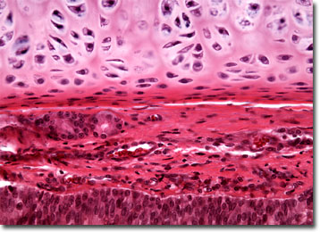
Mammalian Hyaline Cartilage
Cartilage is a tough, but elastic, connective tissue that chiefly comprises the skeletal structures of embryonic and young vertebrates. Sometimes referred to as gristle, the material, which is essentially comprised of a network of collagen fibers embedded in a gelatinous matrix and surrounded by a fibrous membrane called the perichondrium, is usually replaced by the bony skeleton as development proceeds.
In some animals, such as sharks, cartilage remains the chief component of the skeleton even after full maturity is reached. More typical of vertebrates, however, is the permanent retention of small amounts of cartilage in the body. In humans, for instance, the nose, ears, trachea, and larynx are composed of cartilage, and the material can also be found around the joints and in the intervertebral disks. The elastic tissue provides strength, but greater flexibility than bone to such areas and does not contain nerve cells or blood vessels.
Hyaline cartilage is the most prevalent of three types of cartilage found in humans and other vertebrates. It is also the type of the connective tissue that forms the embryonic skeleton. Bluish-gray, pearly, and semitranslucent, hyaline cartilage contains a large number of collagen fibrils dispersed in random directions and a relatively small amount of elastin. It is, therefore, very resilient, but not as flexible as elastic cartilage, which contains significantly greater amounts of elastin. More similar to the composition of hyaline cartilage is fibrocartilage, which also contains large amounts of collagen, albeit in dense bundles that are oriented so that they are parallel to one another.
BACK TO THE BRIGHTFIELD MICROSCOPY IMAGE GALLERY