
Brightfield Microscopy Digital Image Gallery
Clubmoss (Lycopodium) Strobilus
Approximately 200 different species of primitive vascular plants commonly referred to as clubmosses are classified in the genus Lycopodium. The pollen produced by the plants is flammable and was formerly utilized as a flash powder for early cameras and as a common component of fireworks.
View an image of the clubmoss strobilus section at 40x magnification.
Though typically found in modern times creeping along forest floors or tropical mountains, clubmosses were once one of the most prevalent types of plants on Earth. During the Carboniferous period, clubmosses could achieve massive heights, growing as tall as some trees and contributing greatly to the layers of organic material that would eventually develop into the coal deposits commonly used as a source of fuel today. Modern members of Lycopodium are greatly reduced in size from their ancestors, a common variety such as stag’s horn moss only growing about four inches high and ten feet long. Some types are, however, epiphytic and may be seen wrapped around the branches of trees.
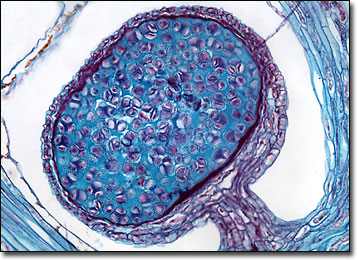
Clubmosses exist in two different forms, although only the aboveground spore producing variety is commonly seen. The leaves of these plants are long and narrow like needles, and strobili, cone-like clusters of diminutive leaves, are usually present as well. At the base of each clubmoss strobilus is a small yellowish-orange sporangium, where the reproductive spores are produced and stored. When conditions are right, the sporangium bursts, releasing the spores, many of which rapidly grow into bisexual gametophytes. In some species, however, the development into the gametophytic form of the plant may take substantially longer, requiring as many as eight years to complete.
BACK TO THE BRIGHTFIELD MICROSCOPY IMAGE GALLERY