
Brightfield Microscopy Digital Image Gallery
Immature Testes
The testes are a pair of male reproductive glands that are sometimes also referred to as the testicles. After human adolescence, the glands are responsible for producing sperm, the male reproductive cells, but before that time remain in an immature state.
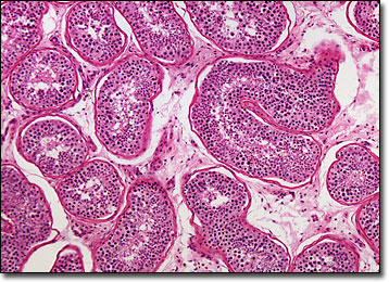
View an image of the immature testes section at 40x magnification.
During embryonic development, the testes originate in the abdomen near the kidneys. Late in the developmental process, around the seventh or eight month of pregnancy, the glands move into their lower position within the scrotal sac. This change of position is regulated by the male sex hormone testosterone, the secretion of which is stimulated by the hormone called chorionic gonadotropin exuded by the placenta. Occasionally, the testes do not descend properly, remaining within the abdomen even after birth. In such cases, medical treatment is usually necessary in order to avoid sterility.
Once the child is born, testosterone production quickly tapers off and stops altogether in a matter of weeks. The resulting lack of the hormone within in the body causes the testes of male children to remain in an undeveloped condition. It is not until the onset of puberty when the pituitary gland begins to produce gonadotropic hormones that sexual maturation of the testes takes place. The hormones secreted by the gland promote tissue growth in the testes, eventually making it possible for the reproductive glands to generate both sperm and androgens.
BACK TO THE BRIGHTFIELD MICROSCOPY IMAGE GALLERY