Brightfield Microscopy Digital Image Gallery
Epididymis
|
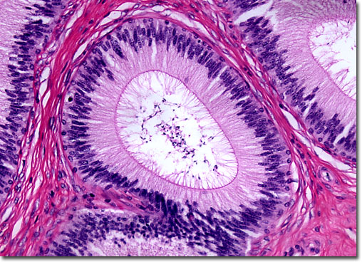
The epididymis, which receives a constant supply of blood from a branch of the testicular artery and is divided into three basic regions, is believed to function in multiple ways. The largest part of the epididymis, known as the head, is located atop the testis, while the smallest part, commonly called the tail, is located where the epididymis separates from the gland. Of intermediate size is the body, which is attached to the rear of the testis and spans its length. The head and body of the epididymis are essential for the maturation of sperm and the tail serves as a storage site for the reproductive cells. The various parts of the epididymis may also be involved in the removal of excess fluid surrounding sperm, but the evidence for this function of the reproductive structure is not conclusive. |
© 1995-2025 by Michael W. Davidson and The Florida State University. All Rights Reserved. No images, graphics, software, scripts, or applets may be reproduced or used in any manner without permission from the copyright holders. Use of this website means you agree to all of the Legal Terms and Conditions set forth by the owners.
This website is maintained by our
|