
Oblique Digital Image Gallery
Mouse Kidney
The kidney is an organ that maintains water balance and expels metabolic wastes in vertebrates and some invertebrates. Primitive and embryonic kidneys have sets of specialized tubules that empty into two collecting ducts that pass urine into a primitive bladder. The more advanced mammalian kidney is a paired compact organ with functional units, called nephrons, that filter the blood, reabsorbing water and nutrients and secreting wastes, producing the final urine.
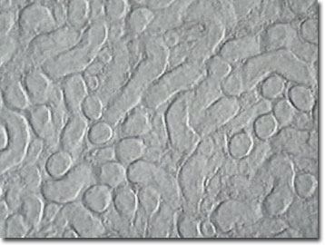
Mouse kidneys are located on the dorsal (upper) wall of the abdominal cavity and are securely held in place by fibrous capsules. Like other mammalian kidneys, the outer layer of the kidney is brownish red and granular in appearance, with a firm consistency. Mouse kidneys are similar to human kidneys, which is why they are often used to simulate human kidneys in scientific studies. Mice have played a significant role in experiments used to diagnose the possible cause and treatment of IgA nephropathy, or Berger's ("burrjays") Disease. This is the most common non-diabetic kidney disease and affects as many as two to four percent of the world's population.
Laboratory mice are special breeds of house mice and are used in many scientific experiments because of their close mammalian relationship to humans. Compared to larger mammals, mice and other rodents are small, easy to handle, inexpensive to house, and breed quickly. During the twentieth century, scientists bred different strains of mice with genetic deficiencies in order to produce models for human diseases.
Contributing Authors
Cynthia D. Kelly, Thomas J. Fellers and Michael W. Davidson - National High Magnetic Field Laboratory, 1800 East Paul Dirac Dr., The Florida State University, Tallahassee, Florida, 32310.
BACK TO THE OBLIQUE IMAGE GALLERY
BACK TO THE DIGITAL IMAGE GALLERIES
Questions or comments? Send us an email.
© 1995-2022 by Michael W. Davidson and The Florida State University. All Rights Reserved. No images, graphics, software, scripts, or applets may be reproduced or used in any manner without permission from the copyright holders. Use of this website means you agree to all of the Legal Terms and Conditions set forth by the owners.
This website is maintained by our
Graphics & Web Programming Team
in collaboration with Optical Microscopy at the
National High Magnetic Field Laboratory.
Last Modification Friday, Nov 13, 2015 at 02:19 PM
Access Count Since September 17, 2002: 11034
Visit the website of our partner in introductory microscopy education:
|
|