Oblique Digital Image Gallery
Insect Cells in Culture
Insect cells, as with other animal cells, feature a cell membrane, nucleus, mitochondria, and other internal organelles and macromolecular assemblies. Unlike plant cells, equipped with environmentally resistant plant walls, insect cells must able to balance internal and external ionic solutions of potassium and sodium. This is particularly problematic for aquatic and wetland species that spend at least a significant portion of their life stages submerged in water, and may partially explain why marine insect species are generally lacking.

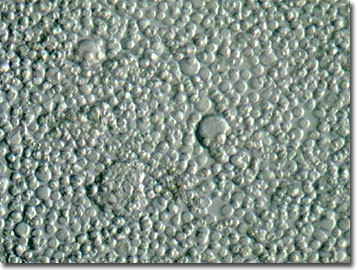
The digital image presented above was captured from a culture of living insect cells with the MIC-D microscope operating in oblique illumination mode. Genetic material, in the form of DNA, is stored and replicated in the nucleus of an insect cell, the "brain" or computer hard drive equivalent of the cell. Mitochondria, the site of power generation via cellular respiration, are responsible for converting ADP to energy-rich ATP while the ribosomes constitute the protein manufacturing centers vital to insect growth and maintenance.
Insect cell lines, such as the ovarian cells of the moth Spodoptera frugiperda, are utilized by histologists and geneticists to study gene expression. They also seek possible causes and therapies for human genetic disorders such as gouty arthritis using insect cell cultures. Foreign DNA is inserted into the insect cells via a virus, and cellular extractions for enzymes or drug trials are then conducted.
Contributing Authors
Cynthia D. Kelly, Thomas J. Fellers and Michael W. Davidson - National High Magnetic Field Laboratory, 1800 East Paul Dirac Dr., The Florida State University, Tallahassee, Florida, 32310.
BACK TO THE OBLIQUE IMAGE GALLERY
BACK TO THE DIGITAL IMAGE GALLERIES
Questions or comments? Send us an email.
© 1995-2022 by Michael W. Davidson and The Florida State University. All Rights Reserved. No images, graphics, software, scripts, or applets may be reproduced or used in any manner without permission from the copyright holders. Use of this website means you agree to all of the Legal Terms and Conditions set forth by the owners.
This website is maintained by our
Graphics & Web Programming Team
in collaboration with Optical Microscopy at the
National High Magnetic Field Laboratory.
Last Modification Friday, Nov 13, 2015 at 02:19 PM
Access Count Since September 17, 2002: 12263
Visit the website of our partner in introductory microscopy education:
|
|
