
Darkfield Digital Image Gallery
Radiolarians
Radiolarians are single-celled protistan marine organisms that distinguish themselves with their unique and intricately detailed glass-like exoskeletons. During their life cycle, radiolarians absorb silicon compounds from their aquatic environment and secrete well-defined geometric networks that comprise a skeleton commonly known as a test.
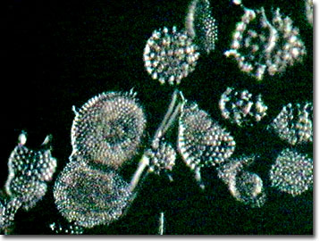
The radiolarian tests are produced in a wide variety of patterns, but most consist of an organized array of spines and holes (or pores) that regulate a network of pseudopods useful in gathering food. When observed with an optical microscope, radiolarian tests are found to be low contrast light-scattering objects that are best viewed using Rheinberg illumination, darkfield illumination, phase contrast, or differential interference contrast (DIC) microscopy techniques. The diversity and beauty of radiolarian tests were first captured and revealed in 1862 by Ernst Haeckel's monograph, Die Radiolarien (Rhizopoda Radiaria), based on specimens gathered from the ocean by the Challenger research cruises of Alexander von Humboldt. The work features 35 exquisite copper plates illustrating hand-drawn radiolarians that still have not been surpassed in quality by modern optical and electron microscopy techniques.
Diatoms, small zooplankton (such as copepods), and other protozoans serve as food sources for the predatory radiolarians. Members of the holoplanktonic radiolaria capture prey items by engulfing them with their pseudopods, a functional characteristic shared by their relatives, the amoebas. The thin, linear ray-form plasmopodia are stretched through the pores of the tests to capture unsuspecting plankton and then retracted when the prey is secured. Digestion occurs in the central cavity. Although most are predatory, radiolaria exhibit wide behavioral variety and include some species that are filter feeders and others believed to be symbiotic with unicellular marine algae (zooxanthellae; more commonly known as dinoflagellates). The dinoflagellate symbionts are enclosed in a thin envelope of cytoplasm produced by the host radiolarian's rhizopodial system. Radiolaria provide ammonium and carbon dioxide for the dinoflagellates, which return the favor by providing the host with a jelly-like layer that serves for both protection and capturing prey. In relationships with marine algal symbionts, the host radiolarian is provided with much needed nourishment when food is scarce, allowing it to survive for several weeks without prey. This type of mutually beneficial relationship is generally found only within radiolarian populations that dwell in water that is shallow enough to receive significant amounts of light (water in the photic zone).
Contributing Authors
Cynthia D. Kelly, Thomas J. Fellers and Michael W. Davidson - National High Magnetic Field Laboratory, 1800 East Paul Dirac Dr., The Florida State University, Tallahassee, Florida, 32310.
BACK TO THE DARKFIELD IMAGE GALLERY
BACK TO THE DIGITAL IMAGE GALLERIES
Questions or comments? Send us an email.
© 1995-2022 by Michael W. Davidson and The Florida State University. All Rights Reserved. No images, graphics, software, scripts, or applets may be reproduced or used in any manner without permission from the copyright holders. Use of this website means you agree to all of the Legal Terms and Conditions set forth by the owners.
This website is maintained by our
Graphics & Web Programming Team
in collaboration with Optical Microscopy at the
National High Magnetic Field Laboratory.
Last Modification Friday, Nov 13, 2015 at 02:19 PM
Access Count Since September 17, 2002: 16596
Visit the website of our partner in introductory microscopy education:
|
|