
Darkfield Digital Image Gallery
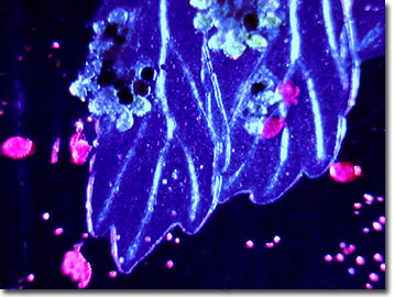
Fern Sori
Fern is a common name for the cryptogamous (spore-producing) plants belonging to the division Filicophyta (also called Filicinophyta). These are primitive vascular plants with true roots, stems, and complex leaves, comprising about 150 genera and as many as 15,000 species.
View a low magnification image of fern sori.
Most ferns reproduce through the alternation of generations, alternating successive generations of sexual and asexual forms. The sexual form, called the gametophyte or prothallia, is a tiny kidney-shaped plant and difficult to find in the wild. The asexual form, or sporophyte, is represented by the fern plant as it is commonly known.
Sporophyte ferns have two methods of asexual reproduction. One is by vegetative cloning, branching off of the root-like underground stem, or rhizome, often forming large, genetically uniform colonies or clones. The second form of asexual reproduction occurs by distribution of spores. These form on the undersides of the leaves in clusters of spore cases called sporangia, or sori (singular, sorus). Sori appear as brown spots and may or may not be present on all leaves. Some species have sori on all the leaves, while others have specialized leaves that bear the sori. When the sporangia dry out, they break open, releasing the spores into the wind. Germination begins when a spore falls in a place with proper conditions of heat and moisture.
Contributing Authors
Cynthia D. Kelly, Thomas J. Fellers and Michael W. Davidson - National High Magnetic Field Laboratory, 1800 East Paul Dirac Dr., The Florida State University, Tallahassee, Florida, 32310.
BACK TO THE DARKFIELD IMAGE GALLERY
BACK TO THE DIGITAL IMAGE GALLERIES
Questions or comments? Send us an email.
© 1995-2022 by Michael W. Davidson and The Florida State University. All Rights Reserved. No images, graphics, software, scripts, or applets may be reproduced or used in any manner without permission from the copyright holders. Use of this website means you agree to all of the Legal Terms and Conditions set forth by the owners.
This website is maintained by our
Graphics & Web Programming Team
in collaboration with Optical Microscopy at the
National High Magnetic Field Laboratory.
Last Modification Friday, Nov 13, 2015 at 02:19 PM
Access Count Since September 17, 2002: 9393
Visit the website of our partner in introductory microscopy education:
|
|