
Brightfield Digital Image Gallery
Trichinella Larvae in Muscle Tissue
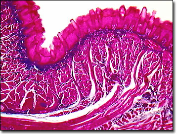
Worm-like helminth parasites are generally classified into two phyla, which are further subdivided into the flatworms (Platyhelminthes) and the roundworms (Nematoda). The digital image presented below was captured with the MIC-D operating in brightfield illumination, and illustrates a stained thin section of human muscle infected with encysted larvae of the Trichinella spiralis roundworm.
Humans are usually infected with the parasite by eating undercooked pork containing the Trichinella larvae, which mature in a few days to a week. Male and female parasite worms mate in the host's intestine, and the female then burrows into the intestinal wall, where she deposits fully developed viable larvae (instead of ova) in the lymph nodes. Larvae traverse the lymphatic system and enter the blood stream, where they are distributed throughout the host and eventually encyst in striated muscle tissue. The most frequently infested muscles are the diaphragm, tongue, biceps, deltoids, and the abdominal walls.
Adult worms, which are 2 to 4 millimeters in length, are barely visible in fecal matter, but can be readily observed with a microscope in the stools of patients during the first few weeks of the infestation. At later stages, identification of Trichinella infestation is made through muscle biopsies and serological testing. The clinical symptoms of Trichinella infestation are mild gastrointestinal disturbances, painful respiration, and muscle pain. Heart muscle damage can ultimately occur.
Contributing Authors
Cynthia D. Kelly, Thomas J. Fellers and Michael W. Davidson - National High Magnetic Field Laboratory, 1800 East Paul Dirac Dr., The Florida State University, Tallahassee, Florida, 32310.
BACK TO THE BRIGHTFIELD IMAGE GALLERY
BACK TO THE DIGITAL IMAGE GALLERIES
Questions or comments? Send us an email.
© 1995-2022 by Michael W. Davidson and The Florida State University. All Rights Reserved. No images, graphics, software, scripts, or applets may be reproduced or used in any manner without permission from the copyright holders. Use of this website means you agree to all of the Legal Terms and Conditions set forth by the owners.
This website is maintained by our
Graphics & Web Programming Team
in collaboration with Optical Microscopy at the
National High Magnetic Field Laboratory.
Last Modification Friday, Nov 13, 2015 at 02:19 PM
Access Count Since September 17, 2002: 17819
Visit the website of our partner in introductory microscopy education:
|
|