
Brightfield Digital Image Gallery
Mallory-Stained Human Tongue Section
Developed by the late Frank Burr Mallory, an American pathologist, Mallory's stain is an important dye used by microscopists and histologists for preparations of connective tissues, fungi and molds, glands and other tissues, such as skin. Although there are several variations on the theme, or the chemical make-up and steps employed, Mallory's stains often feature alum (or iron) hematoxylin, a natural dye that stains nuclei blue and, when coupled with eosin, stains cytoplasm pink.
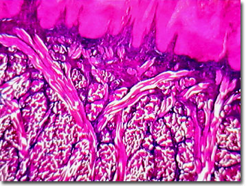
Staining operates by incomplete neutralization of anions by counterions, creating regions of high negative charge density. The high negative charges attract cations, changing the manner in which an anatomical structure or compound absorbs and reflects incident light. For a typical human tongue preparation, Mallory's triple stain is employed. The triple stain contains aniline blue, orange G (OG, an acid dye that stains cytoplasm), and azocarmine (or acid fuchsin). Moderately stained, the collagen fibers of the connective tissues are easily identified by their blue color, while red blood cells inside the blood vessels appear red in color, once dyed. The thin parakeratin layer, which covers the filiform papillae of the human tongue is moderately stained blue. No matter which tissue type is stained with Mallory's triple stain, muscle stains red, as does epithelium, due to red nuclei, while connective tissue in general, stains bright blue. Isolated red blood cells dye to an orange-red when treated with Mallory's trichrome.
Reportedly, Frank Mallory originally experimented with crushed blueberries for a blue vital stain, and later, developed his multiple methods of staining healthy tissue and pathological specimens. Tissue stains are usually classified as acids or bases and their targets as acidophilic ("acid-loving") or basophilic ("base-loving"). Some cellular structures and tissues, referred to as chromophobic, will not absorb stains. As an example, the orange G of Mallory's triple stain is an acid dye that stains cytoplasm and muscle, while eosin has affinity for acidophilic collagen, and hematoxylin for cartilage matrix material. Counterstaining is a technique employed by histologists to reveal nuclei or other features in the background after special stain applications.
Contributing Authors
Cynthia D. Kelly, Thomas J. Fellers and Michael W. Davidson - National High Magnetic Field Laboratory, 1800 East Paul Dirac Dr., The Florida State University, Tallahassee, Florida, 32310.
BACK TO THE BRIGHTFIELD IMAGE GALLERY
BACK TO THE DIGITAL IMAGE GALLERIES
Questions or comments? Send us an email.
© 1995-2022 by Michael W. Davidson and The Florida State University. All Rights Reserved. No images, graphics, software, scripts, or applets may be reproduced or used in any manner without permission from the copyright holders. Use of this website means you agree to all of the Legal Terms and Conditions set forth by the owners.
This website is maintained by our
Graphics & Web Programming Team
in collaboration with Optical Microscopy at the
National High Magnetic Field Laboratory.
Last Modification Friday, Nov 13, 2015 at 02:19 PM
Access Count Since September 17, 2002: 18187
Visit the website of our partner in introductory microscopy education:
|
|