Brightfield Digital Image Gallery
Peyer's Patches
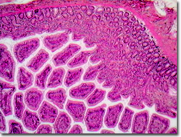
Named for the seventeenth century Swiss anatomist, Hans Conrad Peyer, Peyer's patches are a collection of large oval lymph tissues that are located in the mucus-secreting lining of the human small intestine. These lymph nodules are especially abundant in the ileum, the lowest portion of the small intestine.

Peyer's patches are more numerous in younger individuals and become less prominent with age. Approximately 30 to 40 patches or bundles occur in an individual's intestine, and they appear as elongated thickened areas lacking the villi that are typical of intestinal membrane. However, because they are composed of networking tissues, the nodules are not easily distinguished from surrounding connective tissue.
The lymph system and associated tissues and structures present a strong line of defense against invading bacteria, parasitic microbes, viruses, and other foreign or harmful bodies such as cancer cells. The Peyer's patches contain high concentrations of white blood cells (or lymphocytes) that help protect the body from infection and disease. Because mucus-secreting surfaces of almost any organ, but especially the digestive, genital, and respiratory tracts, are constantly exposed to a wide variety of harmful microorganisms, they are supported by secondary lymph structures. The specialized lymphoid tissues in the small intestine, Peyer's patches, detect antigens such as bacteria and toxins and mobilize highly specialized white blood cells termed B-cells to produce protein structures called antibodies that are designed to attack foreign entities.
Contributing Authors
Cynthia D. Kelly, Thomas J. Fellers and Michael W. Davidson - National High Magnetic Field Laboratory, 1800 East Paul Dirac Dr., The Florida State University, Tallahassee, Florida, 32310.
BACK TO THE BRIGHTFIELD IMAGE GALLERY
BACK TO THE DIGITAL IMAGE GALLERIES
Questions or comments? Send us an email.
© 1995-2022 by Michael W. Davidson and The Florida State University. All Rights Reserved. No images, graphics, software, scripts, or applets may be reproduced or used in any manner without permission from the copyright holders. Use of this website means you agree to all of the Legal Terms and Conditions set forth by the owners.
This website is maintained by our
Graphics & Web Programming Team
in collaboration with Optical Microscopy at the
National High Magnetic Field Laboratory.
Last Modification Friday, Nov 13, 2015 at 02:19 PM
Access Count Since September 17, 2002: 17670
Visit the website of our partner in introductory microscopy education:
|
|
