
Brightfield Digital Image Gallery
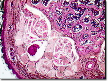
Mouse Tail Thin Section
As recounted in the old nursery rhyme, "Three Blind Mice", the farmer's wife cut off the tails of the mice with a carving knife for running after her, but did she know what she was slicing through? The tail of the mouse is composed of chondroid tissue, bone, tendon, muscle, and skin with hair follicles, as well as nerves and blood vessels.
As a tale of success, the mouse's tail is playing a key role in ongoing stem cell research and development of possible cures for spinal cord injuries. Geneticists favor the tail end of the mouse as a rich source of DNA isolates that are RNA free. The clipped tails of young mice can be successfully stored frozen until DNA extraction is completed, following well-established proteinase digestion protocols, without having to sacrifice the laboratory specimen. Mouse tail is used as a model of human skin in drug trials such as those of topical cream formulations for psoriasis, for cancer research, and is used in other experimental procedures because sampling blood from the tail vein of a mouse or rat is relatively easily carried out.
During embryonic development, approximately ten days following conception, a tail bud appears as a short stump, and then initiates elongation and thinning. The tail of a mouse or rat may be repulsive to some, but to the small rodent, it provides balance, and acts as an effective temperature regulator. As a distinguishing characteristic, the ratio of the relative length of the tail to the body offers rodent taxonomists a method of distinguishing closely related mouse species.
Contributing Authors
Cynthia D. Kelly, Thomas J. Fellers and Michael W. Davidson - National High Magnetic Field Laboratory, 1800 East Paul Dirac Dr., The Florida State University, Tallahassee, Florida, 32310.
BACK TO THE BRIGHTFIELD IMAGE GALLERY
BACK TO THE DIGITAL IMAGE GALLERIES
Questions or comments? Send us an email.
© 1995-2022 by Michael W. Davidson and The Florida State University. All Rights Reserved. No images, graphics, software, scripts, or applets may be reproduced or used in any manner without permission from the copyright holders. Use of this website means you agree to all of the Legal Terms and Conditions set forth by the owners.
This website is maintained by our
Graphics & Web Programming Team
in collaboration with Optical Microscopy at the
National High Magnetic Field Laboratory.
Last Modification Friday, Nov 13, 2015 at 02:19 PM
Access Count Since September 17, 2002: 18713
Visit the website of our partner in introductory microscopy education:
|
|