
Brightfield Digital Image Gallery
Fetal Skull Membranous Bone
The brain of the developing fetus changes so rapidly over the course of gestation that it is possible to date the pregnancy stage by the brain's appearance. Four sutures or bone seams, the coronal (top), lamdiodal (lateral), sagittal, and squamosal (interior) form between the bony plates of the fetal skull as it is developing.
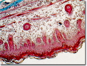
With such rapid change, and because the human brain and head have evolved to progressively larger size, it is important for the fetal skull to be flexible and expandable to protect the brain, but to not interfere with passage through the maternal pelvis at birth. The anterior fontanelle, the point where the sagittal and coronal sutures make contact (known as the bregma in the fetal skull), remains a membranous bone region for about a year-and-a-half after birth, and represents an area of expansion where the sutures are farthest apart. Likewise, the area that develops into the posterior fontanelle in a child, and is termed the lambda in the fetus, initially is composed of membranous bone tissue. As the skull ages, it transforms from membranous bone and ossifies. Membranous bone features active osteoblasts with abundant cytoplasm that secrete the osteoid (non-mineralized bone matrix of Type I collagen). The matrix vesicles (extracellular, membrane-bound fluid-filled cavities) initiate calcification in the osteoid as well as in cartilage and dentin in the embryonic stage of development, and act as the initial nucleation site for mineralization of bone such as skull bones.
Membranous bones include the flat bones of the skull, face, and jaws. These developing head bones form by intramembranous ossification in which embryonic mesenchymal cells differentiate directly into osteoblasts that synthesize the collagenous osteoid. With the addition of calcium phosphate, the membranous bone becomes hard compact bone through the process of mineralization. According to most reconstructive surgeons, membranous bone is superior to endochondral embryologic bone (ribs, iliac, mastoid, sphenoid, and long bones of the skeleton) for bone grafts because it exhibits less resorption. However, membranous bone may take substantially longer to grow than the hollow bones found in limbs, which are based on replacement of temporary cartilaginous skeletal elements with ossified forms.
Contributing Authors
Cynthia D. Kelly, Thomas J. Fellers and Michael W. Davidson - National High Magnetic Field Laboratory, 1800 East Paul Dirac Dr., The Florida State University, Tallahassee, Florida, 32310.
BACK TO THE BRIGHTFIELD IMAGE GALLERY
BACK TO THE DIGITAL IMAGE GALLERIES
Questions or comments? Send us an email.
© 1995-2022 by Michael W. Davidson and The Florida State University. All Rights Reserved. No images, graphics, software, scripts, or applets may be reproduced or used in any manner without permission from the copyright holders. Use of this website means you agree to all of the Legal Terms and Conditions set forth by the owners.
This website is maintained by our
Graphics & Web Programming Team
in collaboration with Optical Microscopy at the
National High Magnetic Field Laboratory.
Last Modification Friday, Nov 13, 2015 at 02:19 PM
Access Count Since September 17, 2002: 18502
Visit the website of our partner in introductory microscopy education:
|
|