
Brightfield Digital Image Gallery
Mammalian Femur
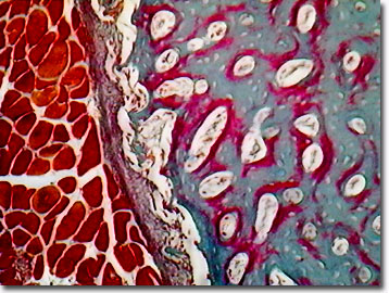
Of the long bones of the mammalian body, the femur or thighbone is the longest, extending from the hip socket to the knee. Long bones are long, cylindrical, have hollow centers, and are characterized by a joint at each end.
View a second image of the mammal femur.
In similarity with the other long bones, the mammalian femur is designed to provide structural support, and various muscles attach to the thighbone to allow movement of the legs. Nerves, veins, and arteries lie along the course of the femur. When the human femur is accidentally broken, the most common treatment involves the application of traction, in which a weight is utilized to place tension on the broken bone to maintain its alignment during healing. The femur joins the tibia (shinbone) and fibula, and in conjunction with the kneecap, or patella, at the knee joint (patello-femoral and tibio-femoral joints), completes the leg skeletal structure.
Scientists have calculated a "strength indicator" value for leg bones as an estimate of various animals' athletic ability. The value represents bone strength in relation to weight. The gigantic femur of the extinct dinosaur, Tyrannosaurus rex, is estimated to have had a strength indicator of "only" 7.5 to 9 square meters per giganewton, where a giganewton is the force needed to move 112,000 tons one meter. In contrast, a runner (faster animals need stronger leg bones), such as the ostrich, boasts a femur strength value of 44 square meters per giganewton, while the muscle-bound mammalian femur of a white rhinoceros measures 26 square meters per giganewton. The relatively small human femur amazingly produces an impressive calculated skeletal strength of 15 square meters per giganewton.
Contributing Authors
Cynthia D. Kelly, Thomas J. Fellers and Michael W. Davidson - National High Magnetic Field Laboratory, 1800 East Paul Dirac Dr., The Florida State University, Tallahassee, Florida, 32310.
BACK TO THE BRIGHTFIELD IMAGE GALLERY
BACK TO THE DIGITAL IMAGE GALLERIES
Questions or comments? Send us an email.
© 1995-2022 by Michael W. Davidson and The Florida State University. All Rights Reserved. No images, graphics, software, scripts, or applets may be reproduced or used in any manner without permission from the copyright holders. Use of this website means you agree to all of the Legal Terms and Conditions set forth by the owners.
This website is maintained by our
Graphics & Web Programming Team
in collaboration with Optical Microscopy at the
National High Magnetic Field Laboratory.
Last Modification Friday, Nov 13, 2015 at 02:19 PM
Access Count Since September 17, 2002: 9937
Visit the website of our partner in introductory microscopy education:
|
|