
Brightfield Digital Image Gallery
Pennate and Centrate Diatoms
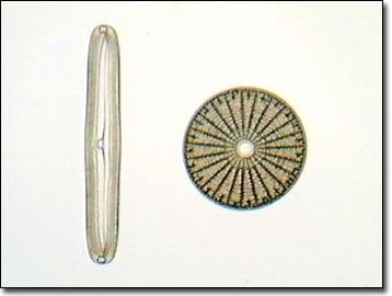
Diatoms have a silicified cell wall forming a pillbox-like shell (frustule) composed of overlapping halves that display intricate and delicate markings useful in testing the resolving power of microscope lenses. The beautiful symmetry and exquisite design of diatom frustules have gained them the title "jewel of the sea."
Depending on the classification system used, diatoms are placed in either the phylum Bacillariophyta, or in the class Bacillariophyceae in the phylum Chrysophyta (golden algae). Most diatoms are planktonic, but some are bottom dwellers or grow on other algae or plants. They are single-celled organisms, although certain types live in extensive colonies. Diatoms are important components of phytoplankton, serving as primary sources of food for zooplankton in both marine and freshwater habitats. Diatomaceous earth, a substance composed of fossil diatom frustules and recovered from deposits formed previously as aquatic sediments, is used in filters, insulation, abrasives, paints, and varnishes.
These organisms usually reproduce asexually by cell division, in which the overlapping shell halves separate, and each secretes a smaller lower half, decreasing in size with each division. Over a period of months there can be as much as a 60% decrease in size. Periodic spore formation serves to restore the diatom line to its original size.
Although diatoms are found in all of the Earth's aquatic environments, most species occur only in habitats with specific physical, chemical, and biological characteristics. Ecologists use this habitat specificity by collecting and analyzing individual species and community data to determine the quality or condition of aquatic habitats. Both long-term monitoring of specific lake and stream habitats and analysis of diatom frustule remains (part of the sedimentary record of lakes) allow scientists to obtain a unique long-term historical perspective on these ecosystems.
Contributing Authors
Cynthia D. Kelly, Thomas J. Fellers and Michael W. Davidson - National High Magnetic Field Laboratory, 1800 East Paul Dirac Dr., The Florida State University, Tallahassee, Florida, 32310.
BACK TO THE BRIGHTFIELD IMAGE GALLERY
BACK TO THE DIGITAL IMAGE GALLERIES
Questions or comments? Send us an email.
© 1995-2022 by Michael W. Davidson and The Florida State University. All Rights Reserved. No images, graphics, software, scripts, or applets may be reproduced or used in any manner without permission from the copyright holders. Use of this website means you agree to all of the Legal Terms and Conditions set forth by the owners.
This website is maintained by our
Graphics & Web Programming Team
in collaboration with Optical Microscopy at the
National High Magnetic Field Laboratory.
Last Modification Friday, Nov 13, 2015 at 02:19 PM
Access Count Since September 17, 2002: 23334
Visit the website of our partner in introductory microscopy education:
|
|