
Brightfield Digital Image Gallery
Goniatitic Cephalopod Fossil
Cephalopods represent a class of mobile predacious carnivore mollusks that possess a bilaterally symmetrical body, a prominent head, and a modified foot composed of tentacles. The digital image featured below was captured from a relatively low-magnification view of a polished thin section from a cephalopod fossil having goniatitic suture morphology.
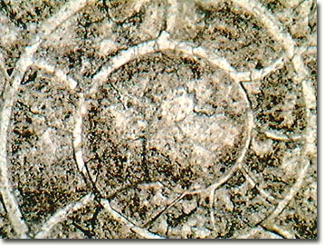
During the Paleozoic and Mesozoic eras, cephalopods achieved a great deal of diversity and abundance in marine habitats, with more than 10,000 species in the fossil record; but only two genera that possess skeletons are known today. Among the modern marine fauna, over 650 species of cephalopods have been cataloged, including squids, cuttlefishes, octopi, and the chambered nautilus. The class also includes the genus Nautilus, which contains the only living cephalopod that retains an external shell.
The cephalopod, whose fossilized shell is presented above, is most likely a member of Ammonoidea, a subclass that evolved from nautiloids during the early Devonian period, about 400 million years ago. These creatures were abundant in the oceans and seas around the world for about 375 million years (as evidenced by the fossil record), but suddenly vanished at the end of the Cretaceous period. The suture structure of the shell has two main features: saddles and lobes, which can be utilized to further classify the fossil. Goniatitic sutures contain lobes that are undivided, ceratitic sutures have serrate lobes, and ammonitic sutures have both lobes and saddles that are finely subdivided.
Contributing Authors
Cynthia D. Kelly, Thomas J. Fellers and Michael W. Davidson - National High Magnetic Field Laboratory, 1800 East Paul Dirac Dr., The Florida State University, Tallahassee, Florida, 32310.
BACK TO THE BRIGHTFIELD IMAGE GALLERY
BACK TO THE DIGITAL IMAGE GALLERIES
Questions or comments? Send us an email.
© 1995-2022 by Michael W. Davidson and The Florida State University. All Rights Reserved. No images, graphics, software, scripts, or applets may be reproduced or used in any manner without permission from the copyright holders. Use of this website means you agree to all of the Legal Terms and Conditions set forth by the owners.
This website is maintained by our
Graphics & Web Programming Team
in collaboration with Optical Microscopy at the
National High Magnetic Field Laboratory.
Last Modification Friday, Nov 13, 2015 at 02:19 PM
Access Count Since September 17, 2002: 12797
Visit the website of our partner in introductory microscopy education:
|
|