
Brightfield Digital Image Gallery
Amphibian Simple Columnar Epithelium
Amphibians, members of the phylum Chordata, are a class of animals that includes the frogs, toads, salamanders, and caecilians. All feature an aquatic larval stage with gills, adult lungs, and external fertilization and development in an aquatic environment.
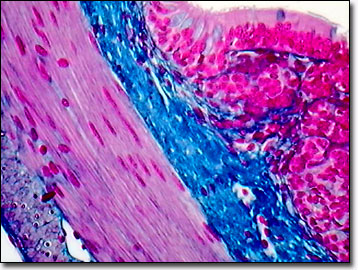
Because some of their life cycle is spent in freshwater ecosystems, amphibians have demands on their skin that terrestrial reptiles and mammals do not. Amphibian skin is permeable to water to allow homeostasis, and to air as a source for respiration. In order to grow, the amphibian must first shed its skin, which also helps control external parasites and may provide some protection as a humidifying cocoon for dry land species during droughts. As a heterotherm or cold-blooded animal, an amphibian's body temperature is partially maintained by fat reserves stored under the skin. In the realm of security, the skin protects against abrasions while color patterns help camouflage amphibians from potential predators. Several species, including the poison arrow frogs and cane toads, feature skin glands that secrete toxins and in some cases, skin pigments and patterns to provide warning coloration (reds, oranges, and yellows contrasted against black) and discourage predation.
The epithelial tissue from amphibians is studied under laboratory controls to assess active transport of water and ion exchange. In the huge role amphibian tissues have played in embryology, the complicated process of gastrulation, including the steps of invagination, ingression, and involution, initiates when the epithelial sheet bends inward, forming an inpocketing. The apical end of the columnar epithelial cells form the central empty space or lumen of the digestive tube during invagination while the basal ends roll inward to form an underlying layer in the involution process. As with the tadpole of the African clawed frog, Xenopus laevis, the intestine is a simple tubular organ consisting of mostly a single layer of primary epithelium. At metamorphosis into an adult frog, the epithelium is drastically remodeled into a complex structure with multiple epithelial folds.
Contributing Authors
Cynthia D. Kelly, Thomas J. Fellers and Michael W. Davidson - National High Magnetic Field Laboratory, 1800 East Paul Dirac Dr., The Florida State University, Tallahassee, Florida, 32310.
BACK TO THE BRIGHTFIELD IMAGE GALLERY
BACK TO THE DIGITAL IMAGE GALLERIES
Questions or comments? Send us an email.
© 1995-2022 by Michael W. Davidson and The Florida State University. All Rights Reserved. No images, graphics, software, scripts, or applets may be reproduced or used in any manner without permission from the copyright holders. Use of this website means you agree to all of the Legal Terms and Conditions set forth by the owners.
This website is maintained by our
Graphics & Web Programming Team
in collaboration with Optical Microscopy at the
National High Magnetic Field Laboratory.
Last Modification Friday, Nov 13, 2015 at 02:19 PM
Access Count Since September 17, 2002: 20602
Visit the website of our partner in introductory microscopy education:
|
|