
QX3 Microscope Light Pathways
The Intel Play QX3 microscope design is similar to the common transmitted light compound laboratory microscope found in universities and hospitals around the world. Illumination from the substage tungsten lamp is diffused when passed through a frosted plastic filter built into the stage where the specimen is placed. This light passes through and around the specimen before entering the front lens of the objective.
Diffusion of the light emitted from the lamp by the plastic screen removes the possibility of seeing details of the filament's structure superimposed over the image of the specimen. In fact, the light emitted by the tungsten bulb in the microscope stage is disorganized and, unlike a standard teaching or research microscope, is not directed into an inverted cone of illumination by a substage condenser or lens system. Instead, the emitted light is first diffused by the mixing chamber before passing through the frosted stage filter and then through the specimen.
The light path from the substage mixing chamber, through the specimen and the objective, and into the active pixel sensor (APS) CMOS is illustrated in Figure 1. After passing through a 19 millimeter aperture opening at the front lens of the 10x objective, light rays are further restricted by a smaller 5 millimeter aperture at the rear of the objective. This light then passes through a dichroic window, which filters unwanted infrared radiation from the light. The dichroic window is positioned within a small plastic enclosure that also contains the APS CMOS chip window.
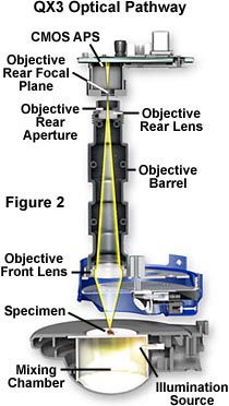
The rear focal plane (illustrated in Figure 2) of the objectives (for the 10x, 60x, and 200x) is also positioned inside the plastic enclosure, which makes this area ideal for insertion of modulation filters, Wollaston prisms, and phase rings for advanced contrast enhancement techniques. Light paths for both transmitted and reflected illumination are similar with the QX3 microscope.
Ray traces of light passing through the Intel QX3 10x objective are illustrated in Figure 2. Light entering the front lens of the objective is focused towards the rear lens and objective rear aperture. The objective rear lens and aperture as well as the rear focal plane are illustrated in Figure 2. The working distance of all three QX3 microscope objectives lies between 26-29 millimeters, although the objectives are not parfocal with one another.
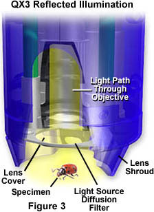
There is also a tungsten lamp positioned in the body of the QX3 microscope near the front lens cover (Figure 3). This lamp is used to provide oblique diffuse reflected light when imaging opaque specimens such as insects, flowers, coins, etc. Light reflected from the surface of opaque objects enters the objective front lens and travels through the microscope as described above (Figures 1 and 2). Auxiliary oblique illumination can also be used to help increase the amount of light reflected from specimens. This is especially important at higher magnifications (60x and 200x) where it is often necessary to increase specimen illumination in order to obtain good images.
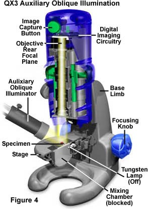
A configuration for auxiliary illumination using an external high-performance tungsten-halide lamp directed by a fiber optic light pipe is illustrated in Figure 4. This is perhaps the best source of external auxiliary illumination that we have found to date. Like oblique illumination from the QX3 body lamp, auxiliary illumination from the light pipe is reflected from the surface of the opaque specimen and enters the objective front lens. After that, the light travels the same pathway described in Figures 1 and 2 (compare Figure 4 to Figure 1). For small specimens, it is advantageous to place a 2-inch square section of dark paper over the stage diffusion filter to avoid transmitted light from the stage from entering the objective during reflected light experiments.
BACK TO INTEL PLAY QX3 MICROSCOPE ANATOMY
Questions or comments? Send us an email.
© 1995-2022 by Michael W. Davidson and The Florida State University. All Rights Reserved. No images, graphics, software, scripts, or applets may be reproduced or used in any manner without permission from the copyright holders. Use of this website means you agree to all of the Legal Terms and Conditions set forth by the owners.
This website is maintained by our
Graphics & Web Programming Team
in collaboration with Optical Microscopy at the
National High Magnetic Field Laboratory.
The QX3 microscope design is copyrighted © 2022 by Mattel, Inc. Intel® Play™ is a registered trademark of the Intel Corporation. These companies reserve all of their rights and privileges under copyright law. The material contained in this website is solely the opinion of the authors and is intended for eduational use only.
Last Modification Friday, Nov 13, 2015 at 02:18 PM
Access Count Since December 24, 1999: 35936
Visit the official Intel Play website:
![]()