
QX3 Digital Image Gallery
Transmitted Brightfield Illumination
Fetal Skull
These thin sections of mammalian fetal skull and flat bones of the face are stained with eosin and hematoxylin. The digital photomicrographs illustrate intramembranous ossification.
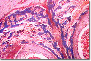

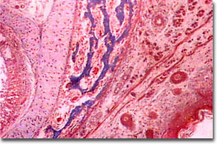
The transmitted brightfield digital images above were recorded using a QX3 microscope that was modified for auxiliary illumination. This was accomplished by directing a fiber optic light pipe at a quarter-inch hole drilled into the front of the mixing chamber to increase the illumination intensity. The light pipe was aimed at the side of the mixing chamber to avoid directing illuminating the frosted diffusion screen.
BACK TO THE BRIGHTFIELD GALLERY
Questions or comments? Send us an email.
© 1995-2022 by Michael W. Davidson and The Florida State University. All Rights Reserved. No images, graphics, software, scripts, or applets may be reproduced or used in any manner without permission from the copyright holders. Use of this website means you agree to all of the Legal Terms and Conditions set forth by the owners.
This website is maintained by our
Graphics & Web Programming Team
in collaboration with Optical Microscopy at the
National High Magnetic Field Laboratory.
The QX3 microscope design is copyrighted © 2022 by Mattel, Inc. Intel® Play™ is a registered trademark of the Intel Corporation. These companies reserve all of their rights and privileges under copyright law. The material contained in this website is solely the opinion of the authors and is intended for eduational use only.
Last Modification Friday, Nov 13, 2015 at 02:19 PM
Access Count Since December 24, 1999: 18126
Visit the official Intel Play website:
![]()