
Advanced Condenser Systems
Achromatic Condenser Improvement
of QX3 Digital Images
The following images compare performance of the Intel Play QX3 Computer Microscope with and without the aid of an organized cone of illumination from a substage condenser containing an aperture diaphragm. These photomicrographs are unretouched and were captured with the QX3 interactive software.
Trinitrotoluene (TNT) in Polarized Light
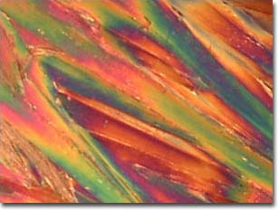 |
| QX3 with mixing chamber (stock - 60x objective) |
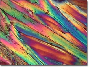 |
| QX3 with achromatic condenser (60x objective) |
Both photomicrographs illustrate crystallites of trinitrotoluene (TNT) photographed with the 60x objective under illumination with polarized light and a full-wave retardation plate. The image on the top was recorded with a stock QX3 microscope after adding the polarizer and analyzer. On the bottom is an image recorded with the QX3 body with the assistance of an achromatic substage condenser (Nikon) using the same components in the optical pathway.
DDT (Pesticide) in Polarized Light
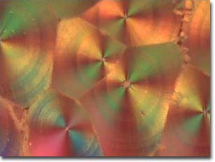 |
| QX3 with mixing chamber (stock - 60x objective) |
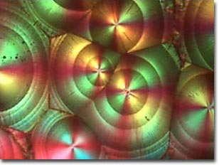 |
| QX3 with achromatic condenser (60x objective) |
Both images above are polarized light photomicrographs of crystallites of DDT (the powerful, but now banned pesticide) with the addition of a full-wave retardation plate between the specimen and the analyzer. The image on top was recorded with a stock QX3 microscope after adding the polarizer, analyzer and retardation plate. On the bottom is an image recorded with the QX3 body with the assistance of an achromatic substage condenser (Nikon) using the same components in the optical pathway.
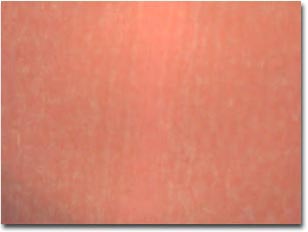
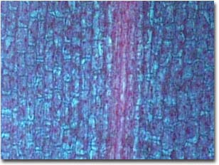
Tilia (Basswood) Stem Stained Thin Section
 |
| QX3 with mixing chamber (stock - 60x objective) |
 |
| QX3 with achromatic condenser (60x objective) |
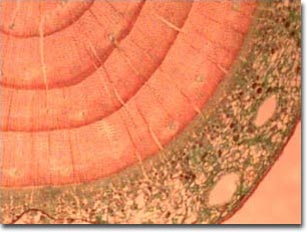
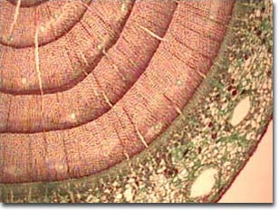
The photomicrographs above were made with a stained thin section of a stem from a basswood tree (Tilia) stained with a quadruple stain and covered with a coverslip. The image on top was recorded with a stock QX3 microscope straight out of the box. On the bottom is an image recorded with the QX3 body with the assistance of an achromatic substage condenser (Nikon) using the same components in the optical pathway.
Blue-Green Algae
 |
| QX3 with mixing chamber (stock - 200x objective) |
 |
| QX3 with achromatic condenser (200x objective) |
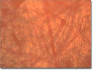
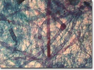
A whole mount of assorted blue-green algae were photographed using the QX3 microscope body with and without the aid of an auxiliary achromatic condenser. The upper image, lacking contrast and color saturation, was made using the microscope in a stock configuration with the mixing chamber. When the condenser is used to provide an even cone of illumination, contrast and color saturation are increased and the image quality is greatly improved, as illustrated in the lower digital image.
Elodea Leaf Tip
 |
| QX3 with mixing chamber (stock - 200x objective) |
 |
| QX3 with achromatic condenser (200x objective) |
The images above are high magnification views of a thin section of Elodea leaf tip stained with a quadruple stain consisting of safranin O (nuclei, chromosomes, and cell walls stain red), fast green (cytoplasm stains green), crystal violet (starch stains blue/purple), and orange G (acidophilic cytoplasm and cell walls stain green). The photomicrographs are intended to illustrate cell and leaf development and were obtained with the 200x objective. Note the detail present in the lower photomicrograph where a substage condenser is used to organize the illuminating light rays. Alternatively, without a condenser (upper image), there is a complete lack of specimen detail.
Additional examples of digital image enhancement through the use of a substage condenser with the QX3 computer microscope are linked below.
Biotin (Vitamin H) Crystallites - The dramatic enhancement of image quality is readily apparent by comparing images of recrystallized vitamin H (biotin) using an advanced achromatic condenser to images obtained with a stock QX3 microscope.
Insect Wing - Notoriously difficult to image in transmitted brightfield illumination, even with a research-grade microscope, insect wings show reasonable contrast and image sharpness with the QX3 microscope when assisted with a substage condenser.
Mosquito Whole Mount - An unstained whole mount of a mosquito is imaged with and without the assistance of an achromatic substage condenser using the QX3 microscope body and interactive software.
Palatine Tonsil Stained Thin Section - A thin section of human palatine tonsil stained with eosin and hematoxylin is imaged with and without the assistance of an achromatic substage condenser.
Recrystallized Silver Nitrate - Enhanced image sharpness and resolution are quite evident in these polarized light digital images of silver nitrate when an auxiliary condenser is used to control illumination.
Recrystallized Urea - Crystallites of the simple organic chemical urea are compared with and without the assistance of a substage condenser.
BACK TO ADVANCED CONDENSER SYSTEMS
Questions or comments? Send us an email.
© 1995-2022 by Michael W. Davidson and The Florida State University. All Rights Reserved. No images, graphics, software, scripts, or applets may be reproduced or used in any manner without permission from the copyright holders. Use of this website means you agree to all of the Legal Terms and Conditions set forth by the owners.
This website is maintained by our
Graphics & Web Programming Team
in collaboration with Optical Microscopy at the
National High Magnetic Field Laboratory.
The QX3 microscope design is copyrighted © 2022 by Mattel, Inc. Intel® Play™ is a registered trademark of the Intel Corporation. These companies reserve all of their rights and privileges under copyright law. The material contained in this website is solely the opinion of the authors and is intended for eduational use only.
Last Modification Friday, Nov 13, 2015 at 02:18 PM
Access Count Since April 24, 2000: 19286
Visit the official Intel Play website:
![]()