Linen Tester Simple Loupe-Style Microscope (circa 1830)


Galleria
License Info
Image Use
Custom Photos
Partners
Site Info
Contact Us
Publications
Home

The Galleries:

Photo Gallery
Silicon Zoo
Pharmaceuticals
Chip Shots
Phytochemicals
DNA Gallery
Microscapes
Vitamins
Amino Acids
Birthstones
Religion Collection
Pesticides
BeerShots
Cocktail Collection
Screen Savers
Win Wallpaper
Mac Wallpaper
Movie Gallery
 

Spike (M.I.) Walker

Mixed Diatoms

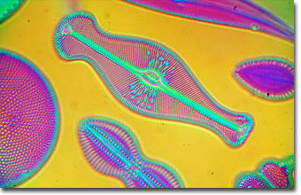
English photomicrographer Spike (M.I.) Walker has been a consistent winner of the Nikon Small World competition for many years and has published many articles and a book about microscopy. Featured below is a photomicrograph of an arranged diatom specimen taken with polarizing interference illumination.

Mixed diatoms

Photomicrograph of mixed diatoms, from an arranged group by Schrader. A Zeiss 10x/0.22 NA polarizing interference objective was used to produce this Jamin-Lebedeff image. Differences in optical wave path are translated into color differences. Illumination was provided by a 12-volt 100-watt lamp utilizing a Zeiss Ultraphot IIIB microscope with 35 millimeter automatic photohead. Photographed with Fujichrome Velvia. (63x)

Diatoms are single-celled algae of the phylum Bacillariophyta and are found in all the Earth's oceans. One of the most prolific sea organisms, the nearly 16,000 species of this group form a significant portion of the aquatic food chain. Diatom cell walls are composed of silica fashioned into a myriad of beautiful geometrical shapes and patterns. Diatomaceous earth, a substance composed of fossil diatoms, is used for making filters, insulation, abrasives, paints, and varnishes.

BACK TO SPIKE WALKER GALLERY

Questions or comments? Send us an email.
Text and graphics for this article are
© 2000-2022 by Spike (M. I.) Walker.
All Rights Reserved under copyright law.
© 1995-2022 by Michael W. Davidson and The Florida State University. All Rights Reserved. No images, graphics, software, scripts, or applets may be reproduced or used in any manner without permission from the copyright holders. Use of this website means you agree to all of the Legal Terms and Conditions set forth by the owners.
This website is maintained by our
Graphics & Web Programming Team
in collaboration with Optical Microscopy at the
National High Magnetic Field Laboratory.
Last modification: Friday, Nov 13, 2015 at 02:18 PM
Access Count Since November 18, 2000: 20323
Microscopes provided by:
Visit the Nikon website. Visit the Olympus Microscopy Resource Center website.