
Spike (M.I.) Walker
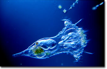
Rotifer (Synchaeta grandis)
English photomicrographer Spike (M.I.) Walker has been a consistent winner of the Nikon Small World competition for many years and has published many articles and a book about microscopy. Featured below is a photomicrograph of a living rotifer specimen taken with DIC illumination.
|
Photomicrograph of Synchaeta grandis. This rotifer was photographed during a field course, using a 16x/0.35 NA planachromat objective and Nomarski DIC condenser on a Zeiss GFL stand. A Zeiss microflash unit was used to obtain the 0.5 millisecond exposure. Photographed with Fujichrome Velvia. (50x) |
Discovered in the late 1600s by Antoni van Leeuwenhoek, rotifers were originally called "wheel animalcules" or wheel animals because their coronas look like turning wheels. This appearance is caused by rippling (metachronal) waves of tiny beating cilia that draw food into their mouths and provide a means of locomotion. Rotifers are the smallest multicellular animals and occur worldwide in primarily freshwater habitats. Nearly all rotifers have chitinous jaws called trophi that grind and shred food. The trophi are the only part of a rotifer that can be fossilized and have been found in amber dating back to the Eocene epoch (38-55 million years ago).
Many rotifer species have no males, females producing only females (parthenogenesis). In some rotifer species, stress can cause females to produce eggs that hatch as males. They have no mouth or digestive tract and die within hours or days. The appearance of males is followed by sexual reproduction. The females then produce resting eggs, which settle to the bottom to hatch when conditions permit. The tiny eggs can withstand desiccation for considerable periods of time and can be carried by wind or birds to any place that holds water (even bird baths and gutters) where they will hatch.