
Wim van Egmond
Micrasterias
Unable to be seen by the naked eye, the microscope has the power to reveal the beauty of the unicellular green algae known as desmids. These tiny organisms exhibit tremendous variability in their cell shape and ornamentation, making them an extremely interesting group to study.
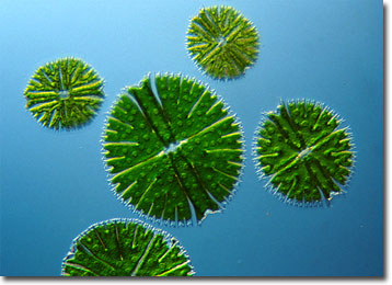
Micrasterias is a genus whose members are considered placoderm desmids, those that feature a cell wall composed of two sections that attach in the mid-region. This type of desmid is also characteristically furnished with pores, spines, granules, or other protuberances. Contrariwise, saccoderm desmids typically possess a smooth, unornamented cell wall that consists of a single piece. As placoderm desmids, the more than 40 known species of Micrasterias exhibit a substantial amount of diversity in the adornment of their lobed, disclike shapes, and many are among the most picturesque microscopic life forms in the world. Most often found in acidic waters and bogs, the organisms may grow between 80 to 200 micrometers in diameter.
The majority of desmids are constricted in the center of the cell, creating two symmetrical semicells connected by an isthmus. Generally, located within the isthmus is the nucleus, and inside each semicell are one or more pyrenoids and chloroplasts. The chloroplasts enable photosynthesis and the pyrenoids are involved in the storage of starch. Reproduction of desmids may involve the sexual act of conjugation or may take place asexually, individuals dividing along the isthmus and each half developing a new semicell. The organisms seem to thrive best in locations that are nutrient poor.
BACK TO WIM VAN EGMOND GALLERY