Batonnet Alignment by High Magnetic Fields
|
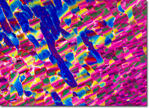
During controlled drying experiments, batonnets formed at the interface between cholesteric and high density liquid crystalline DNA are oriented with respect to externally applied magnetic field direction vectors. Illustrated in the photomicrograph above is a transition region filled with a confluent monolayer of batonnets that are highly oriented. The magnetic field B field vector lies coaxially with the longitudinal plane of the batonnets. The magnification is approximately 250x, and the image was recorded on Fujichrome 64T transparency film using a Nikon Optiphot-Pol microscope with crossed polarized illumination. Exposures were recorded about 3 f-steps under the recommended value given by an in-camera photomultiplier and were push-processed approximately 1.5 f-steps in the first E-6 developer. |
© 1995-2025 by Michael W. Davidson and The Florida State University. All Rights Reserved. No images, graphics, software, scripts, or applets may be reproduced or used in any manner without permission from the copyright holders. Use of this website means you agree to all of the Legal Terms and Conditions set forth by the owners.
This website is maintained by our
|