Observing Mitosis with Fluorescence Microscopy
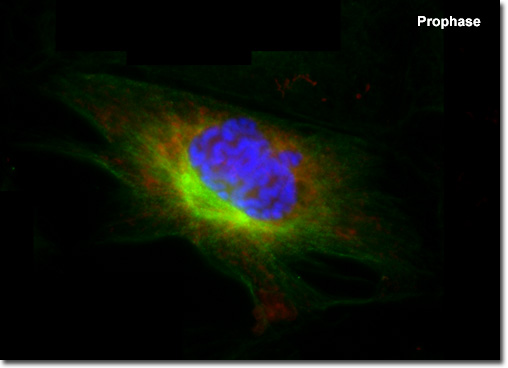
Prophase
|
Presented in the digital fluorescence microscopy image above is a single rat kangaroo (PtK2) kidney cell in the early stages of prophase. The chromatin is stained with a blue fluorescent probe (DAPI), while the microtubule network (mitotic spindle) is stained green (Alexa Fluor 488) and cellular mitochondria are stained with a red dye (MitoTracker Red CMXRos). Each duplicated chromosome contains two identical sister chromatids joined together along their length, with a constricted region occurring at a specific DNA sequence known as the centromere. The chromatin in the centromere exhibits a somewhat higher degree of condensation than the rest of the chromosome. Centromeres are necessary for proper segregation of the sister chromatids during later stages of mitosis. |
© 1995-2022 by Michael W. Davidson and The Florida State University. All Rights Reserved. No images, graphics, software, scripts, or applets may be reproduced or used in any manner without permission from the copyright holders. Use of this website means you agree to all of the Legal Terms and Conditions set forth by the owners.
This website is maintained by our
|