Observing Mitosis with Fluorescence Microscopy
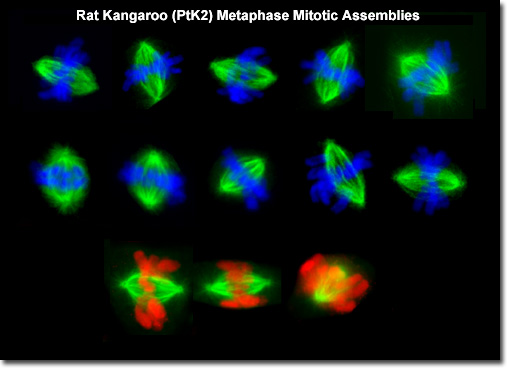
Metaphase
|
Presented in the digital image above is a composite of mitotic rat kangaroo cells showing only the spindle assembly and chromosomes. Fibers comprising the mitotic spindle are functionally divided into two species. The polar fibers extend to the center of the spindle pole towards the metaphase plate, while the chromosomal fibers travel from individual condensed chromosomes to the poles. Chromosomal fibers (also referred to as kinetochore fibers) are attached to the chromosomes at the kinetochores, which form on opposite sides of the centromere. During metaphase, the chromosomes are gradually aligned at the metaphase plate by the mitotic spindle. |
© 1995-2022 by Michael W. Davidson and The Florida State University. All Rights Reserved. No images, graphics, software, scripts, or applets may be reproduced or used in any manner without permission from the copyright holders. Use of this website means you agree to all of the Legal Terms and Conditions set forth by the owners.
This website is maintained by our
|