Observing Mitosis with Fluorescence Microscopy
Anaphase
|
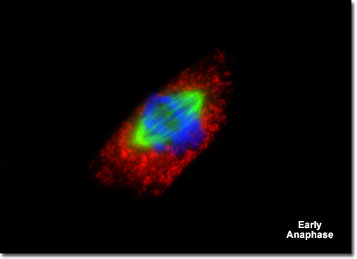
Presented in the digital fluorescence micrograph illustrated above is a single rat kangaroo (PtK2) kidney epithelial cell in the early stages of anaphase. The chromatin is stained with a blue fluorescent probe (DAPI), while the microtubule network (mitotic spindle) is stained green (Alexa Fluor 488) and cellular mitochondria are stained with a red dye (MitoTracker Red CMXRos). The paired centromeres from each chromosome have just begun to separate and the sister chromatids are being pulled apart by the kinetochore microtubules as they are shortened by the loss of tubulin monomers. Migration of the chromatids to opposite poles of the mitotic spindle occurs at a rate of approximately one micrometer per second in most animal cells. |
© 1995-2022 by Michael W. Davidson and The Florida State University. All Rights Reserved. No images, graphics, software, scripts, or applets may be reproduced or used in any manner without permission from the copyright holders. Use of this website means you agree to all of the Legal Terms and Conditions set forth by the owners.
This website is maintained by our
|