Observing Mitosis with Fluorescence Microscopy
Stages of Anaphase
|
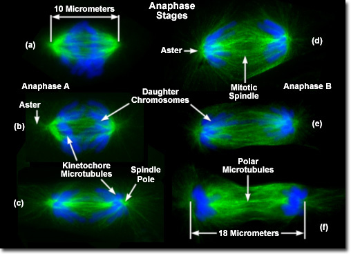
Presented in the digital image above are the mitotic spindles from several rat kangaroo cells (PtK2) in various stages of anaphase. In each case, the condensed chromosomes are stained with a blue fluorescent probe (DAPI), while the microtubule network (mitotic spindle) is stained green (Alexa Fluor 488). Anaphase is characterized by two distinct processes to separate the sister chromatids and move them to opposite spindle poles in preparation for cell division. The first process, termed anaphase A (or early anaphase), occurs with the shortening of the kinetochore microtubules to translocate the chromatids away from the metaphase plate and towards the spindle poles (as illustrated in (a) and (b) above). Anaphase B (or late anaphase) is manifested by a further separation of the spindle poles brought about by lengthening of the polar microtubule network ((c) through (f) above). The overall contribution of each process to the final separation of the sister chromatids varies depending upon the organism being studied. In animal cells, anaphase B begins soon after the chromatids split and terminates when the spindle has grown to approximately twice the metaphase length. In contrast, the cells of many lower eukaryotes exhibit a much larger separation distance during late anaphase, which can result in a spindle length increase up to 15 times the metaphase size. |
© 1995-2025 by Michael W. Davidson and The Florida State University. All Rights Reserved. No images, graphics, software, scripts, or applets may be reproduced or used in any manner without permission from the copyright holders. Use of this website means you agree to all of the Legal Terms and Conditions set forth by the owners.
This website is maintained by our
|